Saturday, 7 May 2016

COUGH AND SNEEZE REFLEX


By 
Tanlaka Lucas Mengnjo
Scientific Journal Club
Faculty of Health Sciences(FHS)
University of Bamenda (UBa),Cameroon 
tanlakalucas@gmail.com


Cough reflex
The bronchi and trachea are so sensitive to light touch that slight amounts of foreign matter or other
causes of irritation initiate the cough reflex. The larynx and carina (the point where the trachea divides into the primary bronchi), are especially sensitive and the terminal bronchioles and even the alveoli are sensitive to corrosive chemical stimuli such as sulfur dioxide gas or chlorine gas.

Afferent nerve impulses pass from the respiratory passages mainly through the vagus nerves to the
medulla of the brain. There, an automatic sequence of events is triggered by the neuronal circuits of the medulla causing the following effects:

First, up to 2.5 liters of air are rapidly inspired.
Second, the epiglottis closes and the vocal cords shut tightly to entrap the air within the lungs. 
Third, the abdominal muscles contract forcefully, pushing against the diaphragm while other expiratory muscles, such as the internal intercostals, also contract forcefully. Consequently, the pressure in the lungs rises rapidly to as much as 100 mm Hg or more. 
Fourth, the vocal cords and the epiglottis suddenly open widely so that air
under this high pressure in the lungs explodes outward.

Indeed, sometimes, this air is expelled at velocities ranging from 75 to 100 miles per hour. Importantly, the strong compression of the lungs collapses the bronchi and trachea by causing their noncartilaginous parts to invaginate inward so that the exploding air actually passes through bronchial and tracheal slits. The rapidly moving air usually carries with it any foreign matter that
is present in the bronchi or trachea. The pathway is shown below:


Sneeze Reflex
The sneeze reflex is very much like the cough reflex except for the fact that it applies to the nasal passageways instead of the lower respiratory passages. The initiating stimulus of the sneeze reflex is irritation in the nasal passageways; the afferent impulses pass in the fifth cranial nerve to the medulla where the reflex is triggered. Series of reactions similar to those of the cough reflex take place. However, the uvula is depressed so that large amounts of air pass rapidly through the nose thus
helping to clear the nasal passages of foreign matter. This is shown in the figure below                                                                                                            


References :TETBOOK OF MEDICAL PHYSIOLOGY ,Eleventh edition by Guyton & Hall,Page 840,Colour ATLAS OF HUMAN PHYSIOLOGY  5th Edition completely revised and expanded,page 133


TRAIN OFF YOUR WEIGHT OR DIET OFF YOUR WEIGHT?

By
CHEKWI  BENWIE
MEDICINE 1
SCIENTIFIC JOURNAL CLUB
Faculty of Health Sciences
University of Bamenda, Cameroon

TANLAKA  LUCAS MENGNJO
MEDICINE 2
SCIENTIFIC JOURNAL CLUB
Faculty of Health Sciences
University of Bamenda, Cameroon
tanlakalucas@gmail.com

TRYING TO LOOSE WEIGHT BY DIET ALTERATION
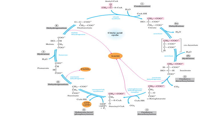
Human beings are often posed with several tragedies among which we have the assertion that it is easier to gain weight than to lose it. In order to detect the cause of this reality, we will take a closer look at the TCA cycle, particularly, the decarboxylation reactions.

The surplus of the food (carbohydrates, proteins and lipids) we consume can be stored as triacylglycerols in adipose tissue. Also these biomolecules are interconvertible except fats which cannot be converted to carbohydrates. Fats gain their way into  the citric acid cycle through the formation of acetyl CoA fed into the TCA pathway by β oxidation of fatty acids. For this to happen, acetyl CoA fed into the TCA pathway will have to yield oxaloacetate, the main precursor for gluconeogenesis. However, the two carbon atoms influxed into this pathway are lost in 2 decarboxylation reactions to produce CO2. Regardless of which compound owned the two carbon atoms, a two carbon loss is a two carbon loss. This results in the disparity in anabolic and catabolic pathway as in the diagram below.


As we have already seen above, fats cannot yield carbohydrates. Glycemia (blood sugar level) is a very critical physiological condition which needs to be regulated. This is because the brain uses gluocose  as its main metabolic fuel. To a lesser extent, like in the case of extreme starvation, the brain can switch to using acetoacetate (ketone body) as a fuel. This can be problematic because ketone bodies are fats and are obviously derived from a nearby source; the meningeal covering of the brain. Therefore, much of our metabolism is tilted towards protecting the master of the body (brain).

The surplus food we consume is stored as fats. If we choose to limit our calorie intake (place ourselves on a diet), the fat stores are mobilized to produce energy. This can lead weight loss. To maintain the normal glycemic level, our bodies release glucagon and adrenaline to breakdown glycogen to produce glucose (glycogenolysis in liver).

Unfortunately, we eventually become hypoglycemic. this is because our glycogen reserves run out since there is little or no intake of carbohydrates. Prior to prolonged hypoglycemia, Growth Hormone and Cortisol are also released. They both decrease glucose utilization by most cells including brain cells. Consequently we become depressed, sluggish and easily irritated. We then start conceiving negative thoughts like "this diet issue is completely pointless, a piece of chocolate cake won't kill me". If we consider our diet, our body now switches to using proteins to maintain blood glucose level. Proteins are converted to pyruvate after being degraded to amino acids .  Hence, we lose muscle as we lose fat.

LOOSING   WEIGHT  BY  EXERCISE
Nonetheless, there is a better and easier way of losing weight, that is, excersise . Appropriate excersice can train our body to use all the fat stores in adipose tissues to supply the tricarboxylic cycle with acetyl CoA through beta oxidation. If we maintain our normal blood glucose level, we save our proteins and we have enough carbohydrates to maintain our glycogen stores.  With a balanced ratio of exercise to food intake (balance intake of nutrients), our fat degradation can be increased without sacrificing our carbohydrate or proteins.Sample applications are illustrated by  the following figure


The  figure above shows a woman in the initial state with extended and/or deformed  abdomen (image on the left ). The image on the right shows this same woman who regained normal size, weight  and form by  simply constantly doing exercise without necessarily restricting her diet. 

Adopt  the "training-off weight"  method  of life ,regain and conserve your normal weight and form.
  Therefore it is easier and healthier to train off weight than to diet it off. It is now left for us to take our stand depending on what we have seen biochemically.


References 
BIOCHEMISTRY International student edition 6th edition
Campbell /FARRELL . chap 19 biochemical connections page 572
GUYTON AND HALL textbook of medical physiology . 12th edition chapter 78 unit XIV
DAVID L. NELSON and  MICHAIL M. COX, Lehninga   PRINCIPLES OF BIOCHEMISTRY,Fourth Edition,Chapters: 15,16.Pages:536,607 ,Figure:14-10,16-7
Bossip.com/945926/weight-loss-sorcery a  gallery of shockingbefore and after waist training photos


Monday, 2 May 2016

Relationship Between Microcephaly and the Zika Virus.


by
Ngouagna Edwin Longchi 
Medical Laboratory Science, 
Scientific Journal Club
Faculty of Health Sciences
University of Bamenda, Cameroon.
edwinlongchingouagna@gmail.com

Content
•Introduction.
•Microcephaly and related causes.
•Zika virus and associated diseases.
•Case study of Brazil, is there a link between microcephaly and the zika virus? , what piece of work is currently going to prove the relationship?, can a vaccine be developed?
•Prevention.
•Conclusion.

Introduction.
What is microcephaly?
Microcephaly is a medical condition in which the brain does not develop properly resulting in a small than normal head. It can be present at birth or develop in the few years after birth. Infants with microcephaly are born either with a reduced or normal head size. Subsequently, the head fails to grow, while the face continue to grow at normal rate producing a child with a small head, a receding forehead, a loose, often wrinkled scalp.

Fig 1: microcephaly (left), normocephaly (right).
People with microcephaly were described as “pin heads”, “monkey man”, and “little boy”.

Causes of microcephaly.
Microcephaly is congenital and acquired.
Congenital microcephaly is caused by gene deletion, single gene defect, autosomal recessive disorder, chromosomal abnormality, or toxin induction during the period of cell migration.
Acquired microcephaly is caused by disruptive injury e.g. ischemic stroke, vertically transmitted infection e.g. toxoplasmosis, others are trauma, metabolic disorders, drugs.
Infants with microcephaly have delay motor functions and speech, hyperactivity, intellectual disability, convulsions may also occur, clumsiness and spastic quadriplegia (paralysis of both arms and legs).
Microcephaly has no cure.

What is the zika virus?
The zika virus is a non-living microorganism that is capable of replicating only inside a host, member of the family flaviviridae, and genus flavivirus and grouped under group iv positive single stranded RNA. Isolated from the zika forest Uganda in 1947. Transmitted by the mosquitoes Aedes aegypti, Aedes albopictus, sexual intercourse, vertical transmission (mother to child).
This virus causes a wide range of infections cutting across dengue, yellow fever, Japanese encephalitis, and microcephaly though a thought as this piece of work is to find out.
Now, we ask the mighty question, is there a relationship between the zika virus and microcephaly?
              In 2015, zika virus RNA was detected in the amniotic fluid of women whose fetuses had microcephaly indicating that the virus had crossed the placenta and could have caused mother-to-child infection.
           In our case study, we look at Brazil and Rio de Janeiro in February 2016 where in Brazil brain tissues from two new borns with microcephaly who died within twenty hours of birth, placenta, and other tissues of two miscarriages (eleven and thirteen weeks) tested positive for zika virus using reverse transcriptase polymerase chain reaction(RT-PCR). In a cohort study of pregnant women in Rio de Janeiro, zika virus infection was associated with fetal death, placental insufficiency, fetal growth restriction, and central nervous system injury (microcephaly and ventricular calcifications) in twelfth out of forty-two studies using ultra sound.
         According to WHO on the 5th of February 2016, a causal link between the zika virus and microcephaly was strongly suspected but not yet scientifically proven although the microcephaly cases in Brazil are spatio-temporarily associated with the zika outbreak in Brazil as reported by the BBC in early march 2016, more careful investigation and research work need to be done to better understand the relationship.
       While waiting for the research work, can a vaccine against the zika virus be produced?
Vaccines exist for many flaviviruses such vaccines for yellow fever virus, Japanese encephalitis but none for the zika virus. However, report shows that in early march 2016, eighteen companies and institutions internationally are working on a vaccine against the zika meanwhile the WHO experts suggested that the priority should be to develop inactivated and other non-live  vaccines that have a safety profile suitable in pregnant women and those of child bearing age. Nikos Vasilakis of the center of biodefense emerging infectious diseases predicted that it may take two years to develop a vaccine, but 10 to 12 years may be needed before regulators for public use approve an effective zika virus vaccine.

How do you and I prevent the zika virus?
           As of February 2016, the CDC recommends that men who reside in or have travelled to an area of active zika virus transmission such as equatorial Africa, south east Asia who have a pregnant partner should abstain from sexual activity or to correctly use condoms in whatever type of sex for the duration of  pregnancy to avoid infection leading to vertical transmission with possible microcephaly  whereas those with non-pregnant partners may require abstinence or condom use though the CDC did not specify how long these practices should be followed.
          Pregnant women should not travel to zika virus endemic regions, as of January 2016, the CDC and other governments issued travel guidelines and warnings.
          In Colombia, Ecuador and some other countries, women were advised to postpone getting pregnant until more is known about the risk.
Microcephaly has no cure so, how do we manage a microcephalic individual?
 Treat the signs and symptoms to confer relief for example speech therapy could help in delay speech.

Conclusion.
The zika virus could be one of the possible causes of microcephaly however; research is currently going on to affirm this.

References.
Understanding pathophysiology; Huether-McCance Brashers-Rote.
Microcephaly-Wikipedia, the free encyclopedia.
Zika virus-Wikipedia, the free encyclopedia.
BBC report of early march 2016.


TYPHOID FEVER (Salmonella typhi)


by
Ngouagna Edwin Longchi, 
Medical Laboratory Sciences, 
Scientific Journal Club, 
Faculty of Health Sciences, 
University of Bamenda, Cameroon.
edwinlongchingouagna@gmail.com


What is typhoid fever?
Typhoid fever is an infectious disease caused by the rod shaped Gram-negative bacterium Salmonella typhi. It is also known as enteric fever, or commonly typhoid.
Typhoid fever is clinically indistinguishable from paratyphoid fever, which is caused by any of the three types of Salmonella enterica (S. typhi, S. typhimurium, S. enteritidis). Incubation period is 10 to 14 days.

Pathogenesis.
Origin.
Life cycle

Brief Origin.
     In 430BC, a plague which some believe to have been typhoid  killed one-third of the population of Athens, the ancient historian Thucydides contracted the disease but survived to write about the plague.
It was originally  isolated in 1880 by Karl Joseph Erberth.

Life cycle.
It is not known to infect animals (i.e.  It is anthroponostic).
It is a multi-organ pathogen that inhabits the lymphatic tissues of the small intestine, liver, spleen and blood stream of infected humans producing toxins.
Invasion of epithelial cells stimulate the release of proinflammatoy cytokines that stimulate an inflammatory reaction.
The acute inflammatory response causes diarrhea and may lead to ulceration and destruction of the mucosa.

Mode of transmission.
Contaminated food (fecal-oral route).
Water supplies.
Close contact with infected host.
Working or travelling to areas where the illness is endemic.
Having a weakened immune system, then drink salmonella contaminated water.

Chronic carriers.
These are people who still harbor typhoid bacteria and can still contaminate food and water even after receiving antibiotic treatment and proving to be free of symptoms. salmonellae persist in the gall bladder

SIGNS & SYMPTOMS.
First stage.
ž  High fever 39c to 40oc.
ž  Headache.
ž  Poor appetite.
ž  Generalized aches and pains.
ž   weakness, and fatigue.
ž  Diarrhoea.
ž  rash(rose colored spots on the chest and abdomen).
    Second stage.
ž  High fever of aboout 40oc.
ž  Delirium(State of violent mental agitation).
ž  Widal test is positive.
ž  Frequent afternoon fevers.
       Third stage.
ž  Intestinal hemorrhage due to bleeding in congested Payer’s patches.
ž  Intestinal peforation.

LABORATORY DIAGNOSIS.
Isolation of bacterium from an appropriate clinical sample e.g. blood, serum, urine, bone marror aspiration, stool(for gastroenteritis, Gram test is used).
Biochemical test: identification of the genus Salmonella.
Serologic testing (Widal test). Here, a serial dilution is made using serum tested against antigens from representatives Salmonella species. The highest diluted serum with positive result is taken as a titer.
High or rising titer to O antigen (≥ 1:160) suggests active infection
High or rising titer to H antigen (≥ 1:160) suggests past infection or immunization.
High titer to the Vi antigen occurs in some cases
Causes of false positive Widal test
 Malaria infection
 other acute febrile illness
 Poor quality reagent
Causes of false negative Widal test
 Specimen collected after antibiotic administration
 Specimen collected at early stage of diseases
 Technical errors.
 There is also a Slide agglutination test/ Kauffman-White system
Used to identify unknown cultures with known sera and important for preliminary identification
of culture.
Required: Salmonella O and H polyvalent antiserum
Method: Mix known sera with unknown culture on a slide.
 Clumping occurs within a few seconds in positive result

      Faeces, blood, or other specimens can also be cultured on:
 Several Non Selective media e.g. nutrient broth.
 Selective media e.g. bismuth sulfide, favor growth of Salmonella
Enrichment media e.g. selenite F broth, Inhibit replication of normal intestinal flora and
permit replication of salmonella.
Differential media e.g Mac Conkey agar for isolation of lactose non-fermenters.
     Any growth is subsequently subcultured onto various agars.
The biochemical reaction of suspicious colonies are then determined on triple sugar agar and lysine-iron agar and a concrete identification is made.

Treatment.
 For cases,
Chloramphenicol
Fluoroquinolones
3rd generation cephalosporins
 For carriers,
Ampicillin followed by cholecystectomy
Cefixime (recommended for children) 
Cholecystectomy (Surgical removal of the gall bladder) in chronic cases.

Prevention.
Have good drinkable water.
Good food supplies.
Wash hands before and after visiting the toilet.
Wash hands before and after eating.
Government provides good waste management and water purification.
Proper treatment of the sick.

Vaccination.
Two licensed vaccines are used:
  Oral live Ty21a vaccine avirulent mutant strain of S. typhi in high endemic areas
  Injectable acetone-killed S. typhi suspensions.


Figure 1: Water filter.
Hope you  own a water filter if not rush and get one.


REFERENCES
Salmonella- medical microbiology: university of Texas medical branch.
Salmonella typhi enterica, David, V. Pollack.
Medical Bacteriology: Ethiopia public health Abilo Tadesse, Meseret Alem
Kayser, Medical Microbiology © 2005 Thieme.
Typhoid fever Wikipedia.